Under construction
The Role of Smades in Endocardial Cushion Development
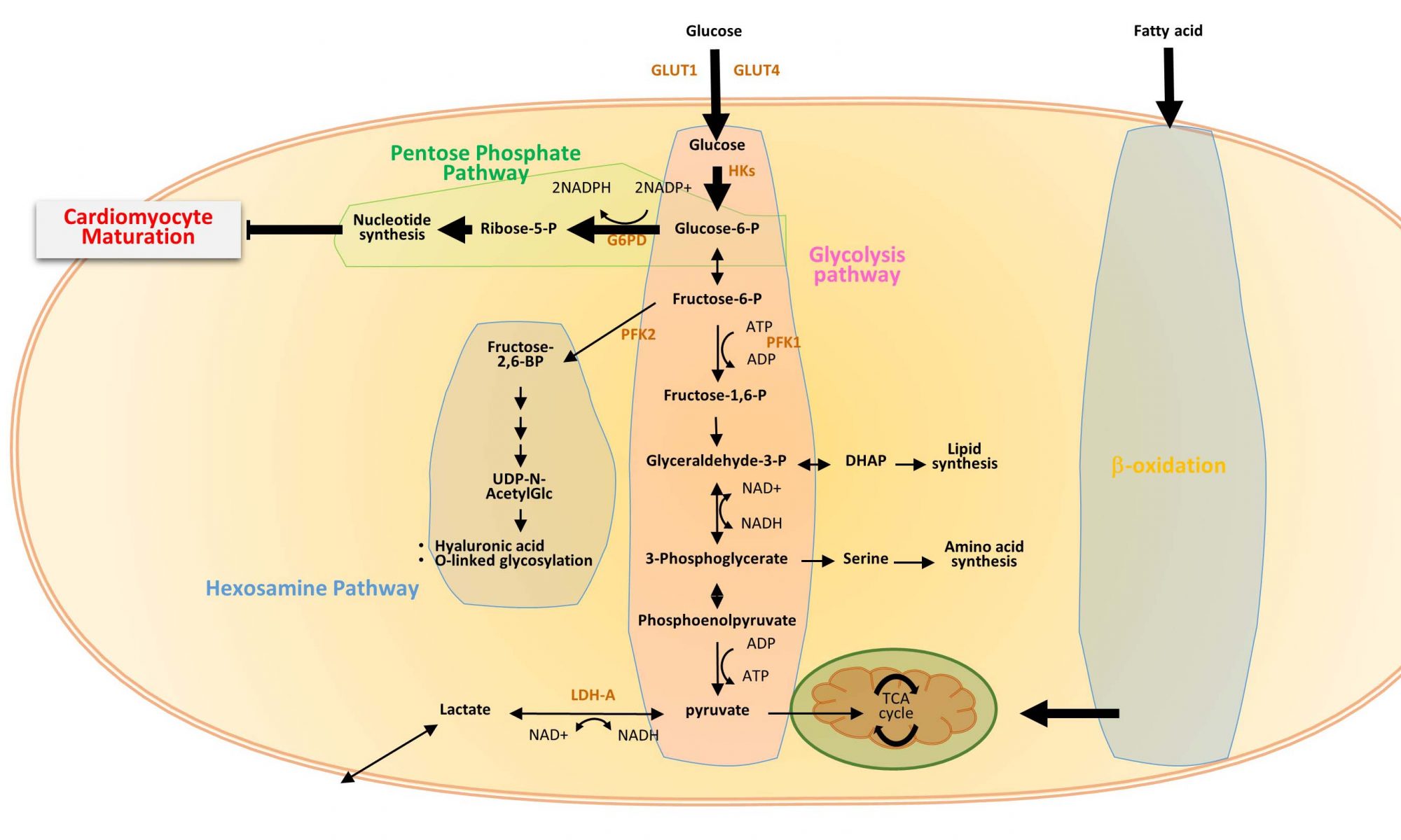
Within the cytoplasm of BMP and TGF-ß receiving cells, Smad6 and Smad7 act as signal attenuators by interfering with Smad4 dependent signal transduction. These inhibitory Smads (iSmads) are structurally similar to other Smad proteins, but lack a C-terminal phosphorylation motif (Whitman 1997). Smad6 mutants on a mixed genetic background exhibit adult outflow tract defects including advanced ossification and cartilaginous metaplasia of the aortic media. The valves of adult mutants also exhibit striking valve hyperplasticity. Given that Smad6 is expressed within the endocardial cushions early in heart development it could play an active role in regulating the number of endocardial cells that undergo EMT and maintaining the proper developmental window in which EMT can occur. Smad6 expression is also maintained throughout development (Galvin et al., 2000), indicating the possibility that it is involved in regulating mesenchymal cell proliferation during valve formation and remodeling. Further, a recent study in the zebrafish model suggests a possible Smad6-mediated mechanism of SHF-progenitor differentiation in the outflow tract (de Pater et al., 2012). However, this has not been addressed within a murine model and could relate to BMP associated congenital OFT defects. We are currently using a global Smad6 knockout allele bred onto a congenic C57/Bl6 genetic background to investigate the role this iSmad plays embryonically in regulating outflow tract and endocardial development. Our initial results show that on a strict congenic background Smad6 knockout results in embryonic valve hyperplasia as early as E12.5. Smad6 mutants also exhibit novel embryonic congenital defects including double outlet right ventricle.
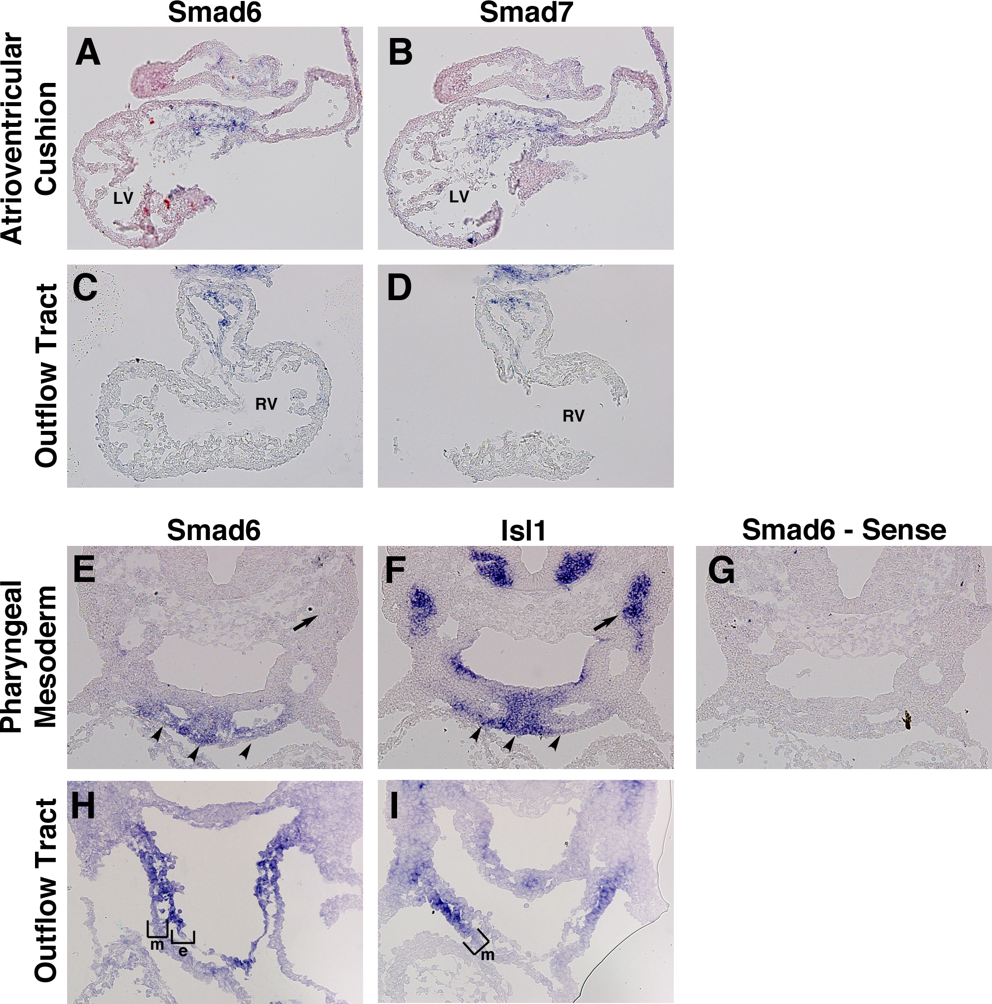
Expression of iSmads during early cardiac development.
In situ hybridization of transverse sections from E10.5 (A-G) and E9.5 (H,I) wildtype embryos. Smad6 and Smad7 have a shared expression domain within both cushion endocardium and mesenchyme of the atrioventricular canal (A,B) and outflow tract (B,C). Smad6 expression co-localizes with the SHF marker Isl1 within the pharyngeal mesoderm (E,F, arrowheads), but not within distal populations of SHF progenitors (E,F, arrows). Control sense reaction demonstrates hybridization specificity (G). Smad6 expression co-localizes with the SHF-marker Isl1 within distal OFT myocardium (H,I, bracket). a – atria, e – endcardium, lv – left ventricle, m – myocardium, rv – right ventricle.