![]() *Please note that all images are Creative Commons CC-BY*
*Please note that all images are Creative Commons CC-BY*
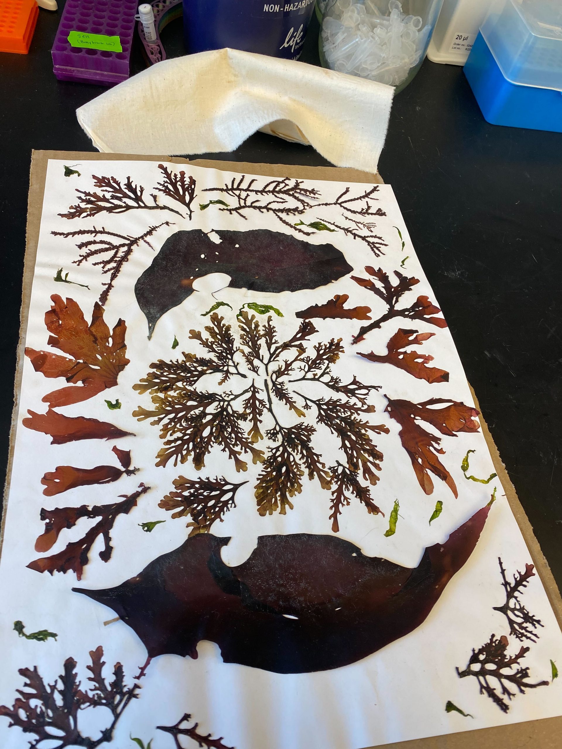
2023.04.17 Practising seaweed ID and pressing skills in Lab Discussion! This beautiful example from Lauren!
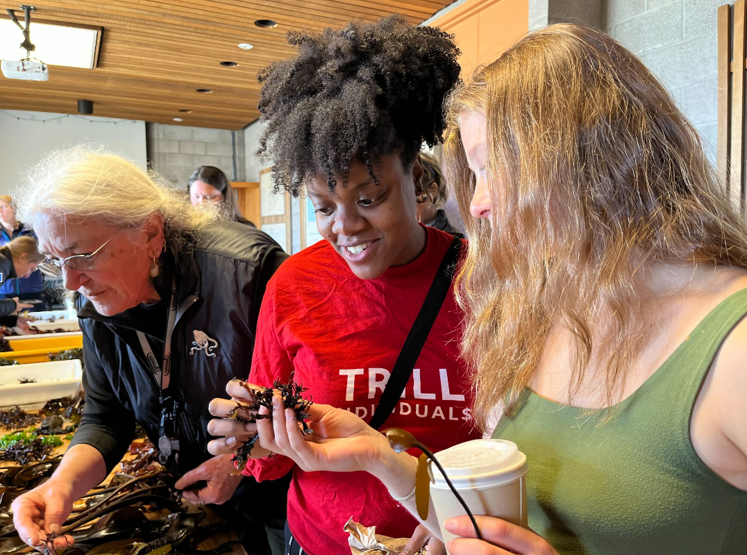
2023.04.14 Learning about seaweed identification from the amazing Dr. Kathy Ann Miller @Bodega Marine Lab (Bodega Bay, CA)
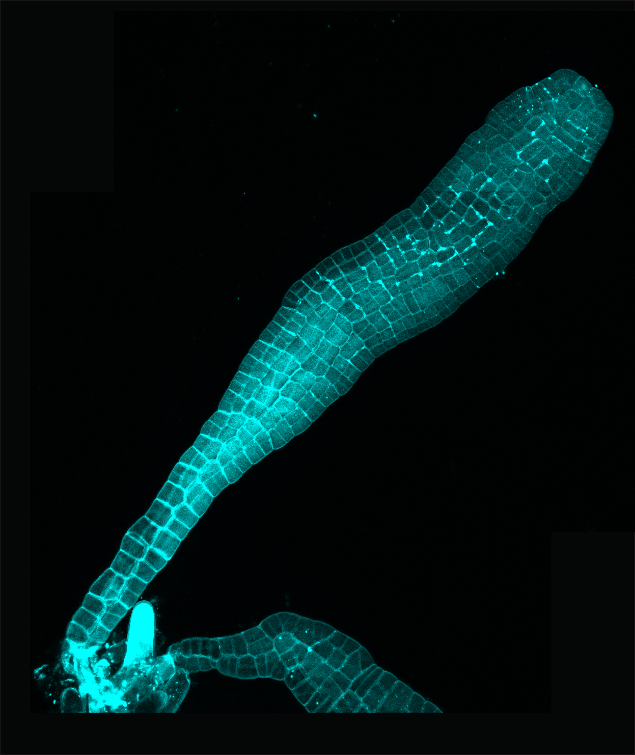
2023.02.01 A giant kelp (Macrocystis pyrifera) embryo, imaged with confocal microscopy using a cell wall stain to outline cells!
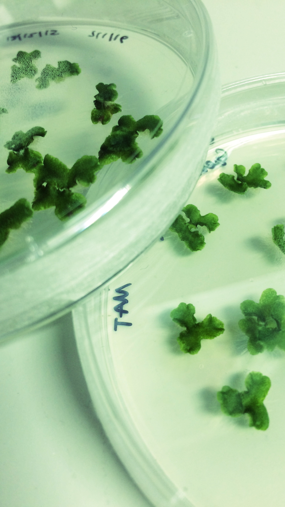
2021.04.29 Seeds received from the wonderful Adrienne Roeder that Chloe & Siobhan were screening for the presence of a fluorescent marker!
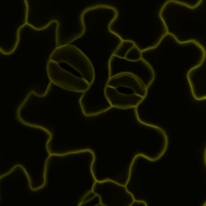
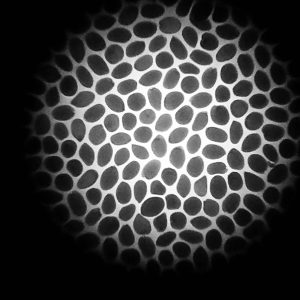
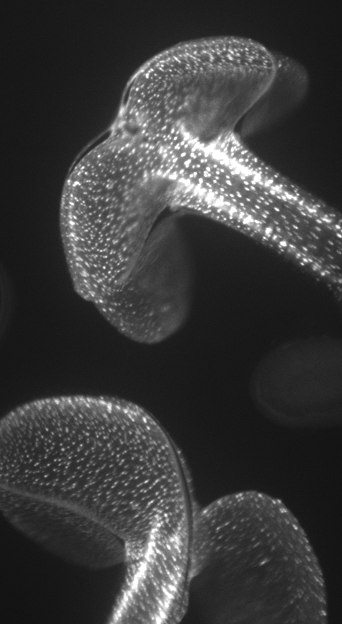
2021.04.08 An adorable epidermal cell arrangement on the Arabidopsis cotyledons surface that we HAD to share! (Even if it is early for Spring…this little chick!)
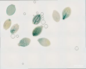
2020.03.24 A beautiful GUS/cotyledon picture to take away quarantine blues.
2020.02.13 Apex of Chara zeylanica, a multicellular green algae. It has a single apical stem cell that produces all of the algal body.
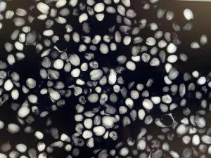
2020.01.08 Arabidopsis seeds seen with an iPhone!
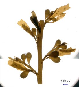
2019.03.13 Sargassum branching….beautiful brown algae!
2019.01.29 Fucus DNA purified using ultracentrifugation and cesium chloride, from Marina!
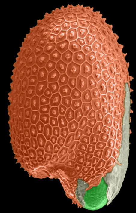
2019.01.01 A new year and a new plant’s life begins! Just germinated Arabidopsis seed, scanning electron micrograph with false colouring, by Firas. Seed coat in orange, endosperm in light green, and the embryo radicle in bright green – bursting through both!
2018.11.16 A graveyard of AFM tips! Had a wonderful day making myself familiar with our new JPK AFM at UCLA 😀
2018.10.16 A gif of epidermal cell peels from our recent NPH paper!
2018.07.17 Accidentally created some art while making a radial tissue map of a hypocotyl!
2017.07.20 Apprently Firas likes building towers in the lab……and is also enjoying screening the MAGIC parent lines for phenotypes. Maybe enjoying is a strong word, but we are grateful for his dedication!

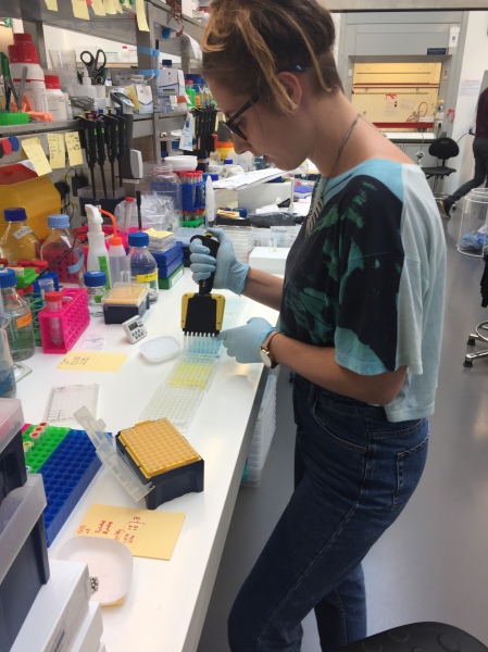
2017.07.18 Marina is learning ELISAs in the lab from Tom…..looks like it is going ok! She also has excellent hair cuts.
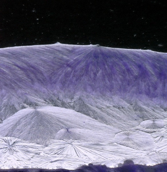
2017.07.07 When you work with mannitol, sometimes you encounter strange new worlds! Firas is the best at documenting them!
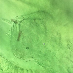
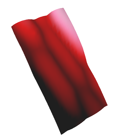
2017.04.11 AFM-based indentation can be applied to organisms other than plants. We have been working on methods to use in macroalgae (methods chapter coming soon!). Below you can see the results from a young Fucus embryo’s rhizoid. The most rigid area (white) is the glass slide it is growing on. The central, elongated, area is the rhizoid which is surrounded by mucilage. The square is 100um wide and high.

2017.04.10 The Easter run-up has us thinking a lot about eggs…..Fucus eggs! We are approaching our last harvest, but hopefully we can eek out a few more fertilisations.

2017.03.10 Searching for the puzzle piece…..these epidermal cell shapes are canonical to modern plant developmental and cell biologists. But how common are they? Stay tuned to find out…..

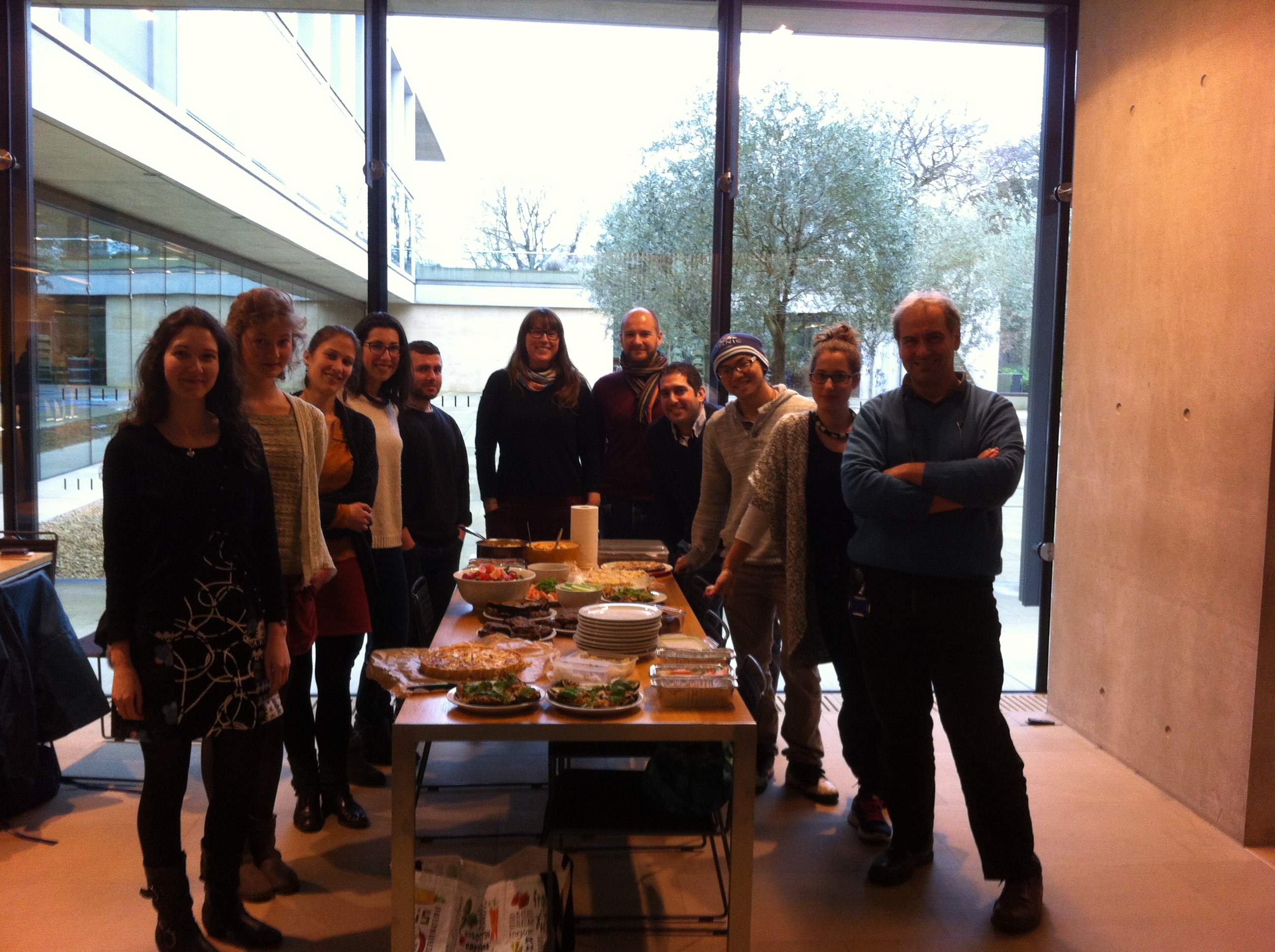
2016.12.12 Holiday Lunch at King’s College. What a great bunch of people we have!

2016.12.05 Happy Holidays from The Braybrook Group. Our tree serves up some Dynasty-class with a Joan Collins angel.

2016.08.19 Dynamics in a single cell: Microtubule movement in a hypocotyl cell by Firas, forward advancing ends are labelled with GFP-END BINDING PROTEIN1.
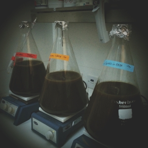
2016.07.20 Jack and Tom are hard at work: working even when they have gone home, seaweed extractions don’t sleep!
2016.07.20 Visiting G. Charras at UCL today with Louis, see evidence of the masters at work (poor N. benthamiana!). Undertaking some never-before-undertaken AFM work.
2016.06.30 Summer lab potluck 2016!

2016.06.02 Marco has been making things! Micro-electrophysiology here we come…

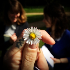
2016.05.09 Wandering around the Botanic Garden, mapping out the Phyllotaxis Tour for Festival of Plants with Katie and Joanna.
2016.03.04 This week we prepared our first RNAseq libraries….a lot of pipetting, but soon we will see what happens, courtesy of the SLCU MySeq.

2016.02.24 An evening confocal session seems a bit more like a journey through outer space. Preparing to finally discover what happens to a plant cell under load…it is actually a far-out project 😀

2016.02.22 Joanna was screening for epidermally expressed nuclear markers, and she made art in the process! They look like they are dancing…..
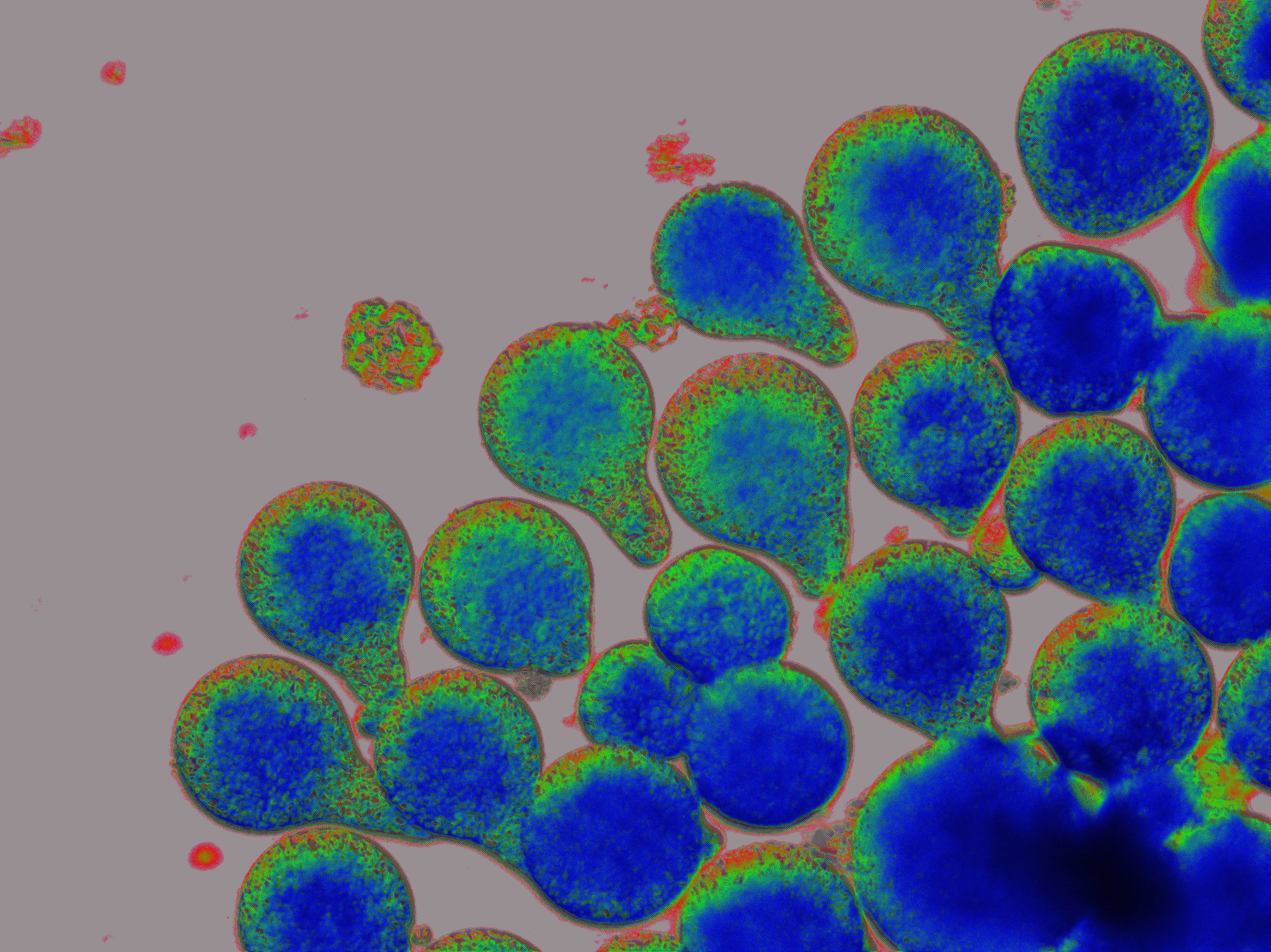
2016.02.03 Fucus zygotes, in technicolor! Marina is tracking cell divisions in Fucus embryos, and this is the beautiful result of her most recent attempts:

2016.02.02 It’s a team effort in the Braybrook Group: harvesting over 2500 hypocotyls, these two are amazing! (they barely needed The Boss, kept shooing me away…)
2016.01.28 The Gemmaelings are coming! Marchantia has taken metaphorical root in the lab. Watch this space….
2016.01.18 January Lab Potluck and Projects Review.
2016.01.16 After a long delay, the Fucus season has begun!
2015.11.22 Another sunny, but chilly, harvest day in Rottingdean. Marina scours the rocks for female Fucus…

2015.11.19 Lab meeting, where Firas tells us all about the hypocotyl!

2015.11.09 There is an Engineer loose in the lab! Louis is learning some biology with Joanna (note: lab coat!).

2015.10.06 Autumn colour comes to SLCU and The Botanic Garden. Here is Rozi out sampling leaves!

2015.09.27 Team Algae pearticipated in ‘The Great Seaweed Harvest of 2015’ wherein we drove to Rottingdean and collected tons of brown algae for our experiments! And it happens to have been a beautiful day…..

2015.09.17 Old school cut-outs! Imaging epidermis cell shapes in diverse species means getting the scalpel out and getting creative with some old shcool protocols. Smoke bush on Gingkgo.
 2015.09.10 A new start. Two first leaves in an Arabidopsis seedling marked by newly formed trichomes. Confocal image of cell membranes (green) and chloroplasts (pink) by FB.

2015.08.20 Hunting for brown algae (a Cornwall holiday). Spent some time on the rocks muttering about brown algae in Cornwall! Meet Fucus vesiculosus, Ascophyllum nodosum, and Fucus serratus :

2015.06.29 New lab pets. There are three new photosynthesising organisms in this tank….can you spot/name them? Hint: we are going Old School.

2015.04.02 Easter egg hunt! We had a team Easter egg hunt today! Thanks Tom 😀

2015.03.31 Is that a MONKEY?! Oh wait….maybe it is just a developing maize tassel. The cool things you see using an SEM.
2015.03.31 Contamination- it happens to everyone….but does anyone else see the beauty in it? Firas does.

2015.03.19 Brachypodium distachyon makes its appearance in the lab. Stay tuned for some exciting experiments from Rozi!

2015.03.06 First in a (hopefully) long line of Hot-Dog-Bet challenges. Tom lost out to Marco on a bet surrounding mechanical inhibition of growth. Tom, you are Top Dog in spirit, if not in the bet!

2015.02.08 Chalk talk in Plant Development class at The University of Cambridge.

2015.02.20 Moss is undeterred by British winter!

Even through flowers are slim in the UK winter months- you can find many interesting plants still thriving! Plantlife.org.uk has a hitlist of cool looking moss, lichen and fungi for you to discover!
2014.11.28 Ginkgo leaf drop!
 Gingko trees prepare their petioles for leaf drop, and as soon as a frost hits- all of the leaves abscise and drop within a 24 hour period. Winter is here!
2014.11.06 Seaweed harvest site: Cromer on the Norfolk coast.

2014.10.31 Lab Science Pumpkin Montage!

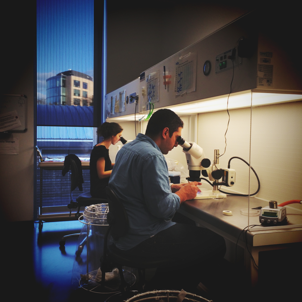
2014.09.30 In the lab again……..what a beautiful place to work.

2014.09.30 Standing up to AFM! These seedlings are poised to be mechanically tested by the AFM……..they look up to the test.

2014.08.22 Lab Lunch and send off for Simon (hopefully temporary!). This may also be our Christmas party……we celebrate summer instead!

2014.08.15 An Elastic Forest. Here, the elastic modulus of an Arabidopsis leaf is plotted in Z against X,Y position.

2014.07.19 Representing plant sciences at the SDB Meeting in Seattle (http://www.sdbonline.org/2014mtg) with the other Siobhan! See more on Siobhan Brady’s research at http://www-plb.ucdavis.edu/labs/brady/

2014.06.30 Why is the Endoplasmic Reticulum called the Endoplasmic Reticulum ? Because it really is reticulated! Reticulated is an adjective that means to be arranged like a net. Check out this confocal image of an Arabidopsis cell taken by Firas which has GFP-labelled ER and ER-bodies:

2014.05.16 In preparation for the Festival of Plants, we have made some nice trichome SEMs….Check out the surface of a venus flytrap! The oragnge hair in the center is the trigger hair, for sensing fly presence. The yellow ones are glands for secreting digestive juices….

2014.05.08 Firas makes some awesome hypocotyl movies!

2014.04.22 Floral Chaos! Deconstructing flowers of a new Outreach activity, with Emily and Firas.

2014.04.17 Tour of The Cambridge University Herbarium (in SLCU!). Darwin’s sample from the Beagle and some amazing drawings by Henslow. http://cambridgeherbarium.org/

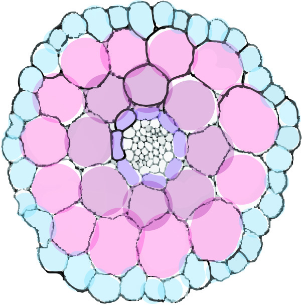
2014.04.01 Assessing cell geometries in the hypocotyl

2014.03.18 Pavement cell rigidity as determined by AFM
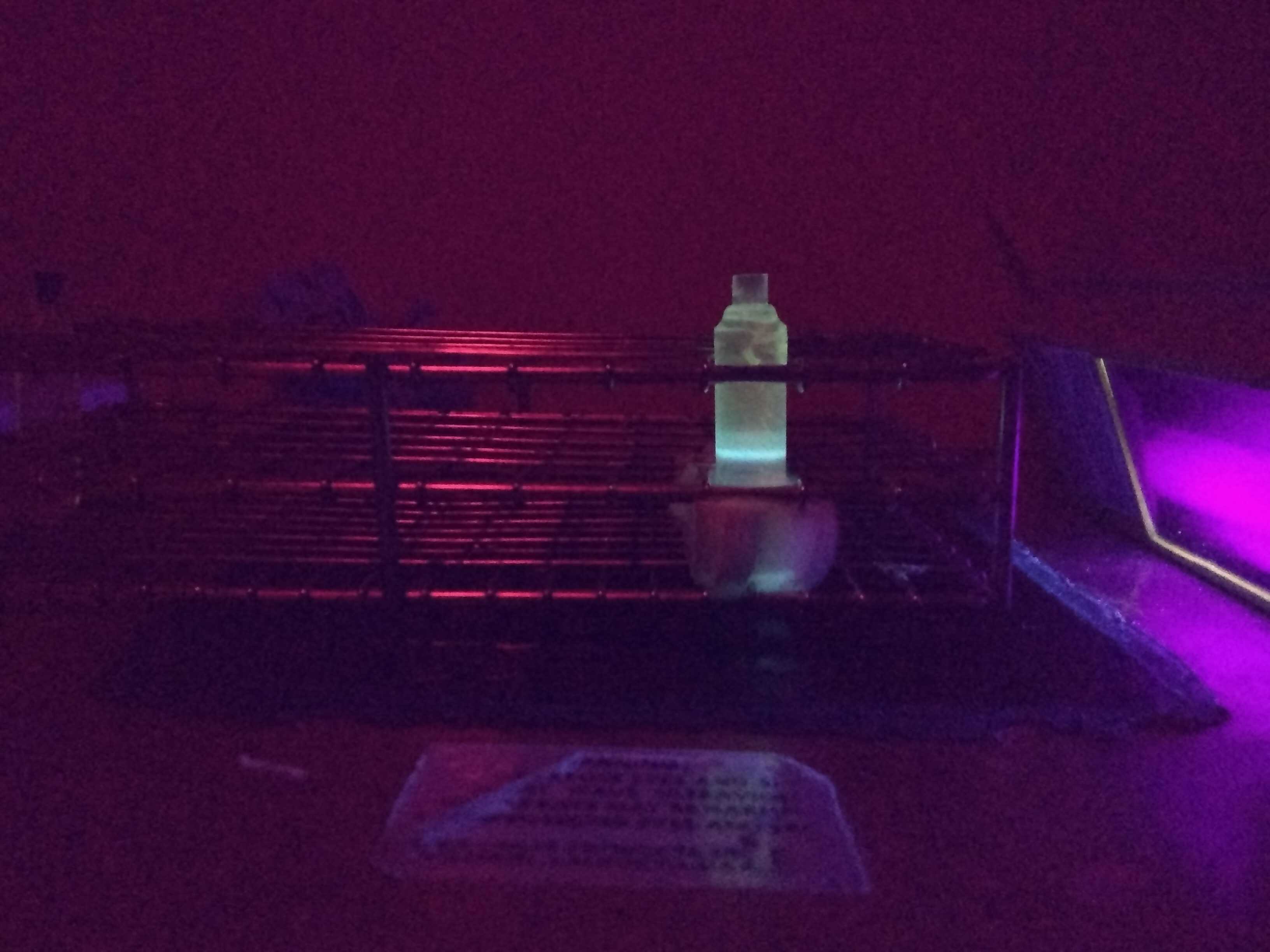
2014.03.11 Infrared cameras are fun……this one was donated by a friend (Tom White, King’s College). A normal digital camera that has ‘night mode’ allowing it to image with IR.


2014.03.05 Gestalt shift, brought to you by the leaf epidermis! This image shows chlorophyll autofluorescence (blue), microtubules (red), and actin (green). And eyes!?!?! By Firas.
2014.02.27 Andy Warhol inspired germinating seeds. ID coloring in top leftt: yellow seed coat, pink endosperm, and blue radicle. It is pretty important to stage germination for hypocotyl research.

2014.02.25 Late night/early morning hypocotyl AFM! We can use the AFM to measure cell wall mechanics, but also to re-create surfaces.
2014.02.20 Teaching some cool kids about the plants they eat!
SLCU Outreach is fun 😀

2014.02.14 Happy Valentine’s Day from us! Image by Firas.

2014.02.11 This is what brown algae microspore prints look like! With our friend Tom Torode from the Knox Lab in Leeds.

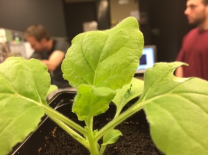
2014.01.30 Arabidopsis seedlings standing tall and reaching up, just like us!

2014.01.14 Imaging set-up No.1 is go!